Pinched Nerve
Pinched Nerve
WHAT IS A PINCHED NERVE
Just like the roots of a tree come together to form the trunk, in the spine nerve roots come together to form nerves in the back which run down through your pelvis then down into your legs. Nerve roots are like cables. Each root contains millions of fibers. Some of the fibers in the nerve root cable are for movement, some are for sensation, others make you sweat. If you pinch a nerve root it hurts!
SYMPTOMS OF A PINCHED NERVE
The pain is sharp, stabbing, and runs down your leg like electricity. Over time damage to the nerve root from compression results in a patch of numbness in the painful leg, as well as weakness. Where you feel it depends on which nerve root is pinched and what that pinched root normally does. That’s how Doctors can predict which nerve root is pinched based on your symptoms and examination. For example, the L5 nerve root has fibers which signal the tibialis anterior muscle to lift the toes when walking, and other fibers which allow you to feel your big toe. Pinching the L5 nerve root causes a sharp, stabbing, electrical pain to shoot form your back down your leg into your big toe; and a foot drop when walking. On examination your doctor finds weakness of the tibialis anterior muscle, the extensor halluces muscle, numbness to your big toe, and normal reflexes. If the weakness is severe, or the pain has gone on for more than 12 weeks, your doctor will confirm the pinched nerve root by MRI.
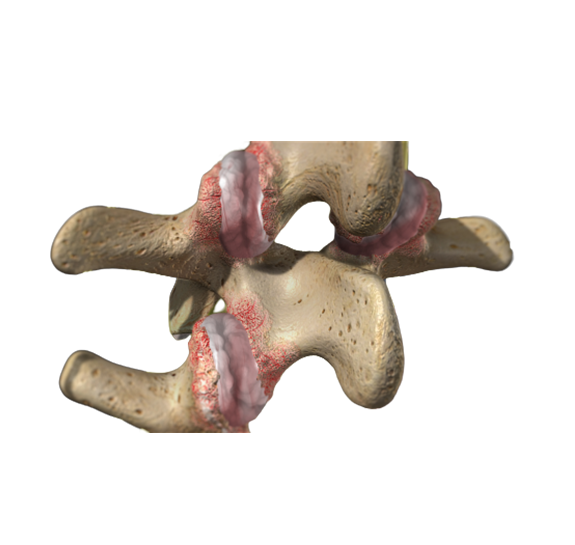
CAUSES OF A PINCHED NERVE
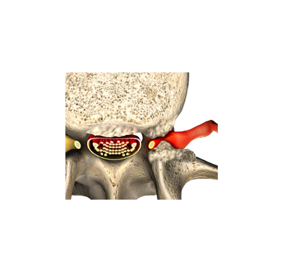
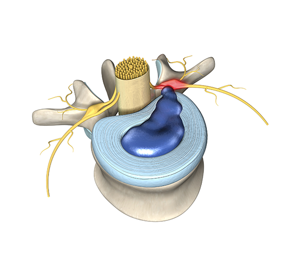
Lots of bad actors pinch nerve roots, and you can see them all on MRI. The most famous one is the Herniated Disc. Disc material is super irritating to nerve roots; the disc doesn’t have to pinch the root much to damage it by irritation. Over time old herniation calcify, and the joints of the spine develop bone spurs which can pinch nerve roots as well. If one spinal bone moves forward on another nerve roots can become trapped, causing severe pinching. MRI shows all of these villains in high definition. If you can’t have an MRI we can still see them in black and white on CT.
APPROACH TO A PINCHED NERVE
We suggest a minimally invasive approach to decompress pinched nerves. A surgeon will start with a small, approximately 1/2 inch incision, in your low back. Using a fluoroscope they place a tubular dilator over the lamina bone, which covers the spinal canal. Operating through a microscope, they shave off a small amount of the lamina bone, retracted nerve root, and then drill out all the bone spurs, also called osteophytes, compressing the nerve root. In most cases the surgical part of the operation takes 30-45 min. They expect their patients wake up with some pain in the back, but without there radiating pain in the leg.
PATIENT EDUCATION VIDEO
ADDITIONAL RESOURCES
Learn more about Herniated Discs which could be pinching a nerve
Explore Nerve Root Pain in the Your Pain Section
Understand what to expect when getting an MRI